23 Mar When a limp isn’t just a limp: How our advanced CT imaging helps diagnose hidden elbow problems in dogs
When a limp isn’t just a limp: How our advanced CT imaging helps diagnose hidden elbow problems in dogs
When your dog develops a limp, it’s natural to assume it’s a minor strain or a sore joint that will improve with rest. But for some dogs, particularly medium and large breeds, intermittent front-leg lameness can signal a more complex orthopaedic issue that standard X-rays may not detect.
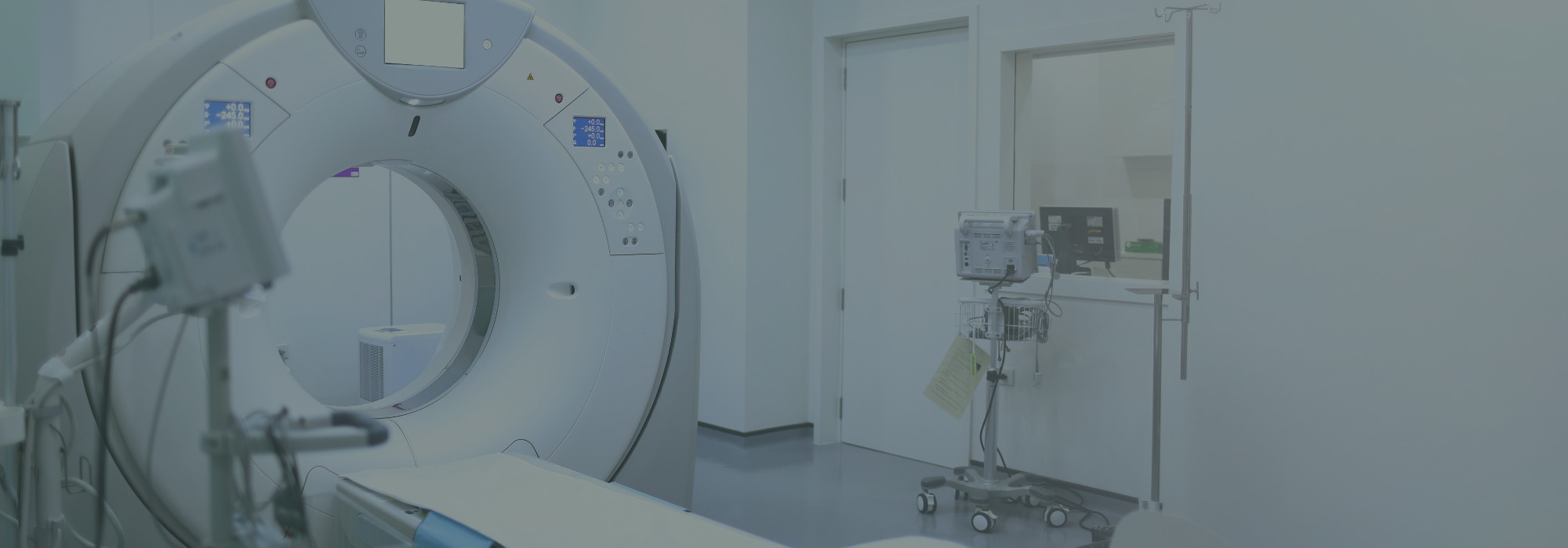
At Wolfe Vets (with practices in Fulham and Chiswick), we use advanced diagnostic imaging, including our on-site CT scanner, to investigate subtle lameness and elbow problems that might otherwise go undiagnosed.
For conditions such as Humeral Intracondylar Fissure (HIF), CT imaging can make a crucial difference in detecting the problem early and helping dogs return to comfortable, active lives.
What Is Humeral Intracondylar Fissure (HIF)?
Humeral Intracondylar Fissure, commonly known as HIF, is a fissure or small crack that develops in the lower portion of the humerus, close to the elbow joint. While the fissure itself may be tiny, it can lead to significant problems if left untreated. Dogs with HIF may experience:
- Intermittent front-leg lameness
- Mild elbow pain or discomfort
- Reduced willingness to exercise
- Stiffness after activity
In some cases, dogs show very subtle symptoms, or none at all, until the bone suddenly fractures during normal activity such as running or jumping. Because of this risk, early diagnosis is extremely important. It can prevent more serious injuries from occurring, as well as improve their overall well-being with an accurate and timely diagnosis.
Dog breeds more commonly affected by HIF
At Wolfe Vets in Fulham and Chiswick, we most commonly see HIF in certain breeds, including:
- Springer Spaniels
- French Bulldogs
- Newfoundlands
- Cocker Spaniels
It is thought that these breeds (and in particular, Spaniels) are more commonly affected due to a combination of lifestyle, genetics and conformation.
However, any dog with persistent or unexplained elbow pain or front-limb lameness should be carefully evaluated by a veterinary professional.
Why elbow problems like HIF don’t always show on X-rays
Traditional X-rays are often the first step when assessing orthopaedic issues in dogs. They provide excellent images of bones and joints, but very small internal cracks, like those seen in HIF, can be difficult or impossible to detect.
This means some dogs may continue to show signs of lameness despite normal-looking radiographs. If symptoms persist, advanced imaging is often the next step to reach a clear diagnosis.
How CT scans help diagnose dog elbow problems
A CT scan (Computed Tomography) provides highly detailed, cross-sectional images of your dog’s bones and joints.
Unlike traditional X-rays, CT imaging allows us to examine the elbow from multiple angles and detect very small structural abnormalities inside the bone. At Wolfe Vets, our advanced CT scanner in London allows us to:
- Detect Humeral Intracondylar Fissures that may be invisible on X-rays
- Evaluate complex elbow joint conditions in dogs
- Diagnose the cause of chronic or intermittent lameness
- Plan orthopaedic surgery with greater accuracy
- Provide faster answers for pet owners
This technology plays a key role in diagnosing conditions affecting active dogs who love to run, jump and play.
Why is early diagnosis of HIF so important?
When HIF is detected early, it may be possible to treat the condition before a complete fracture occurs. Treatment options may include preventative stabilisation surgery, which reinforces the bone and reduces the risk of a catastrophic break during everyday activities.
Early diagnosis can therefore:
- Prevent serious fractures
- Reduce recovery time
- Protect long-term joint health
- Allow dogs to return safely to normal activity
For many dogs, identifying the condition early can truly be life-changing. Our founder, Dr James Bennett, is recognised by the Royal College of Veterinary Surgeons as an Advanced Practitioner in Small Animal Surgery, which, alongside our state-of-the-art equipment and experienced team, means we can carry out a wide range of complex surgical procedures, and in particular, orthopaedics, at our practice.
Advanced veterinary imaging at Wolfe Vets
At Wolfe Vets, we are proud to offer advanced veterinary imaging services for pets across Fulham and Chiswick. Our on-site CT scanner allows our veterinary team to quickly investigate orthopaedic problems, including:
- Persistent or unexplained lameness
- Elbow pain in dogs
- Suspected HIF
- Complex joint disorders
- Sports or activity-related injuries
Having CT imaging available within our practice means we can often reach a diagnosis faster and more accurately, helping your dog receive the most appropriate treatment as soon as possible.
When should your dog have a CT Scan?
Your vet may recommend a CT scan if your dog has:
- Ongoing front-limb lameness
- Elbow pain that does not match X-ray findings
- Recurring stiffness after exercise
- A breed predisposed to HIF
- Orthopaedic problems requiring detailed investigation
For many pet owners, CT imaging provides the answers that traditional diagnostics cannot.
Supporting active pets across London
Pets thrive when they can move freely, play, and explore the world around them. Hidden orthopaedic conditions like HIF can quietly threaten that freedom if they remain undiagnosed.
If your dog is experiencing chronic lameness, elbow pain, or unexplained front-leg discomfort, our veterinary teams in Fulham and Chiswick are here to help investigate further and guide you toward the best possible treatment options. Early answers can make all the difference in keeping your dog happy, active, and pain-free.
Sorry, the comment form is closed at this time.